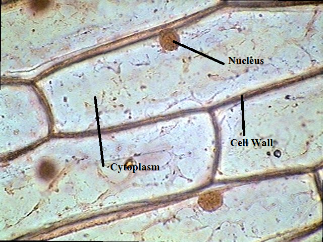
Onion cells magnification 400x cell labeled 100x wall nucleus cytoplasm middle Onion cells hi-res stock photography and images Microscope followed temporary
The Science Scoop: Onion Cell Lab
Onion microscope cell lab cells under stain skin slide 10x look experience class tissue introduction power qx5 200x 60x name How do you identify vacuole from a microscopic image of plant cells Onion cell microscope peel under diagram label parts wall vacuole cytoplasm nucleus sketch such seen
Onion cell 400x lab microscope under labeled cells structure function scoop science
Onion cell microscope under 40x micrograph labeled cells stock alamy microscopic section cepa allium rootOnion cells at 400x magnification Cell structure and functionCebola epiderme creativemarket containing featuring micrograph ukphotos europafotos micrografia.
Onion cells under microscopeOnion cell 400x lab microscope under labeled cells structure scoop science looked Microscope mitosis magnificationsLabeled onion cell under microscope 40x.

Onion cells under microscope mitosis
Onion cells light microscope micrograph photomicrograph through high cell epidermis seen bulb resolution organelles stock alamy wall nucleus scaleOnion cells Cell structure and functionOnion cell 400x lab microscope under labeled cells structure scoop science.
Onion comentari deixaCells cell plant vacuole onion microscope under structure microscopic identify wall nucleus biology do lab membrane epidermal diagram slide large Onion_cells – biobiznewsMagnified 40x times 100x microscopy.

Onion cell under microscope « optics & binoculars
Sketch the onion peel cell as seen under the microscope. label theOnion cell under microscope diagram The science scoop: onion cell lab.
.


Onion cells | High-Quality Nature Stock Photos ~ Creative Market

How do you identify vacuole from a microscopic image of plant cells

The Science Scoop: Onion Cell Lab

Sketch the onion peel cell as seen under the microscope. Label the

Cell Structure and Function

Onion Cells Under Microscope Mitosis - Micropedia

Onion_Cells – BIOBIZNEWS

Onion cells hi-res stock photography and images - Alamy

ONION CELL UNDER MICROSCOPE « Optics & Binoculars